
FluoroStar is the optimal solution for everyday work. It combines performance, maneuverability, modern achievements in obtaining, processing and storing information.
The FluoroStar Digital Mobile Imaging System is characterized by high performance and excellent maneuverability. The system's 1K x 1K or 752 x 582 image processing capabilities, using the latest imaging technologies, produce superb image quality. With flexible and ergonomic mechanical properties, the system contains a touch screen with a user-friendly interface that meets the requirements of surgeons for imaging.
Месяц: Июль 2023
X-RAY DEVICE C-ARCH GENERAL ELECTRIC BRIVO OEC 715 785
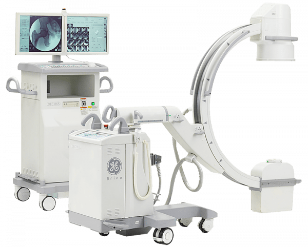
Brivo OEC 715 is the basic solution for orthopedics and traumatology.
Brivo OEC 785 is a device with extended functionality and options.
Advantages:
Obtaining good imaging results is primarily due to positioning accuracy. The Brivo OEC 715 and Brivo OEC 785 GE models are lightweight and easy to move.
The body of the C-shaped apparatus is designed for ease of maneuvering and is flexible enough to move to almost any position. The quick connection feature allows you to start working immediately after moving the machine.
At the generation stage, a specialized X-ray tube developed by GE is used.
It is possible to easily change the parameters of fluoroscopy: the level of radiation intensity is regulated, the choice between four pulsed modes is provided.
The high resolution raster and detector provide a high level of contrast.
At the stage of data processing, the contrast, brightness and clarity of images are optimized, an effective algorithm for removing artifacts is used.
Two swivel professional medical monitors ensure the transmission of visual information without distortion and do not tire the doctor.
Thanks to the ergonomic user interface, learning how to use the system takes a minimum of time.
The machine's information system provides the freedom to choose the place and format of data storage. For storage of pictures CD, DVD, USB are used. DICOM interface will provide integration of the device with medical local networks, connection to network printers and workstations. The total system memory can hold up to 80,000 images. Uninterruptible power supply eliminates equipment failures and loss of patient data.
DENSITOMETER OSTEOSYS MEDONICA DEXXUM-3
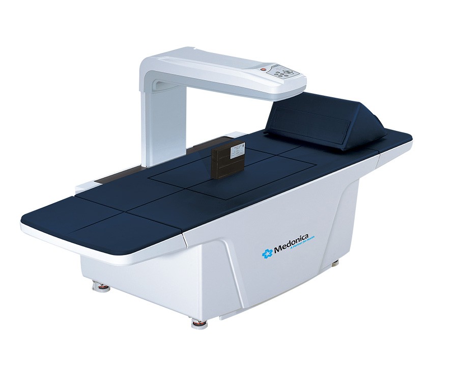
The bone x-ray densitometer DEXXUM-3 allows you to determine bone mineral density using dual-energy absorptiometry, recognized as the "gold standard" for the universal diagnosis of osteoporosis risk, since it combines a number of advantageous qualities: the ability to study the axial skeleton, high sensitivity and specificity, accuracy and reproducibility, low dose x-ray exposure, the speed of research. The design of DEXXUM-3 is specially designed for the convenience of all ages of patients. Its height from the floor is only 64.2 cm, which is especially important for elderly patients. Application area The equipment is intended for examination of the spine and femurs. It is widely used in rheumatology, obstetrics, gynecology, endocrinology and traumatology to detect early changes in the skeletal structure at the preclinical stage, with the aim of further treatment. Peculiarities The high scanning speed of the DEXXUM-3 densitometer allows you to get research results in 85 seconds. for the spine and in 65 sec. for the thigh bones. The continuous scan process is patented and consists of parametric imaging of the skeleton by rearranging a series of 2D images sequentially recorded during linear continuous displacement. The software allows you to sort and compare measurement data by date of examination and by density. DEXXUM-3 is a reliable, multifunctional densitometer for hospitals and clinics. Main characteristics Automatic determination of bone mineral density and display of results in the form of an absolute indicator of BMD (content of mineral density per 1 sq. cm. of the scanned surface), each area of the examined surface. Editing (adding and deleting) the bone scan area allows you to reduce the level of errors in the calculation of the BMD index, especially when scanning with implants, after fractures or operations. Daily quality control (phantom measurement) guarantees high stability of the results. Reproducibility of studies not less than 99% User-friendly software, support for several languages, including Russian, color image of data, customization of reports at the request of the customer. Results display: as BMD (Bone Mineral Density) values in g/cm. sq. in the form of a T-test, the ratio of the patient's actual bone mass to the typical (maximum) bone mass of young healthy patients of the same sex, calculated as a standard deviation (SD) value. in the form of a Z-criterion - the ratio of the patient's bone mass to the average age bone mass of the reference group, calculated as a standard deviation value.
DENSITOMETER GENERAL ELECTRIC LUNAR IDXA

X-ray densitometer.
Allows you to study the anteroposterior projection of the spine, hip, two femurs, femoral neck and much more.

Advanced technology in bone tissue diagnostics and body composition analysis
The allowable patient weight limit is 204 kg;
DICOM interface: worksheet storage, printing and management;
Line detector with high pixel density and direct conversion technology.
In less than 5 minutes and 6 clicks, the Lunar iDXA densitometer can evaluate a patient's bone system. The narrow fan beam satisfies all the needs of the clinic for routine examinations. Thanks to this digital system, the densitometer allows you to perform both standard and more complex studies. The Windows XP-based enCORE workflow uses visual cues to provide reliable results, high throughput, and ease of use. This software allows you to exclude the "human factor" from the process of obtaining information. The auto-analysis function allows you to make accurate calculations in the minimum time, a simple and concise display of the report contains all the information about the patient and an assessment of the risk of bone fracture. The ability to build tables and graphs allows you to evaluate the dynamics of bone density.
MAMMOGRAPH GENERAL ELECTRIC SENOGRAPHE PRISTINA
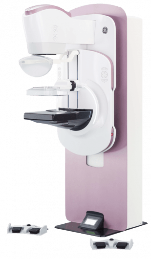
Medical product type code 191110

A full-format digital mammography system that opens up wide opportunities for breast imaging, including in oncology.
The mammographer optimizes exposure settings based on breast tissue properties.

Peculiarities:
Active detector area: 24 x 29 cm;
Tomosynthesis for examining women with a dense breast structure;
Rotation: -180/+180 degrees;
Improved system ergonomics;
The Seno Iris workstation is designed to assist radiologists.
The Senographe Pristina GE mammography system makes it possible to detect breast cancer (BC) at an early stage, which allows you to take screening and diagnostic mammography to a new level of development.
The system of tomosynthesis (3D-mammography) allows using these technologies today as the main method for detecting breast cancer pathologies for a large number of patients, which was not possible for a long period of time.

Breast cancer is one of the most common malignant tumors, which leads to a large number of deaths among women around the world. The use of tomosynthesis technology makes it possible to identify and cure this disease by 30-40%. Experience in medical practice shows that patients who seek treatment at an early stage of cancer are 80% more successful in treatment and recovery compared to those with higher stages.
In oncology, Senographe Pristina offers one of the most sought-after and advanced technologies - CESM contrast spectral mammography. Contrast mammography allows you to detect neoplasms in the mammary glands of any density, to recognize even small (from 5 mm) cancer foci. The technology is highly informative in preparing for surgery and evaluating the effectiveness of breast cancer treatment.
MAMMOGRAPH GENERAL ELECTRIC SENOGRAPHE ESSENTIAL

GE offers a state-of-the-art, reliable solution for mammography, an area where imaging quality is of the utmost importance. The Senographe Essential digital X-ray machine is a system that allows you to perform both routine examinations and advanced diagnostic and interventional procedures. Many additional features are available to expand functionality:
zoom photography,
stereotaxic biopsy,
contrast spectral mammography,
tomosynthesis.
Senograph essential. Diagnostic quality
Digital flat-panel detector based on amorphous silicon 24x31 cm in size, together with the FineView intelligent image sharpening system, it provides the opportunity to obtain maximum diagnostic information.
An automated system for setting exposure parameters allows you to determine the most dense areas of tissue and set radiation parameters and filtering type, providing the best image quality at optimal dose load.
Tissue Equalization is a tissue equalization function that allows you to identify and correct over/underexposed areas in images.
Senograph essential. Performance
Increased throughput is ensured by pre-set automatic collimation, automated tripod positioning, precise adjustment of the angle of its rotation, ease of preparation for the exposure procedure.
Two-speed motor drive increases the speed of patient admission without affecting the accuracy of disease detection
A specialized X-ray tube creates radiation with a spectrum that increases the penetrating power into the breast tissue. The minimally short time between exposures provides favorable conditions for screening.
Senographe Essential.Functionality
Screening and targeted biopsy configurations are available. The GE Healthcare biopsy solution provides precise access to the breast lesion and optimal patient positioning. It allows you to quickly switch from the mode to the mode of interventional procedures and vice versa.
SenoClaire's tomosynthesis feature provides layered tissue imaging. It makes diagnostics even more accurate with a lower dose load compared to standard research methods. This provides excellent sensitivity for morphological disorders and neoplasms, ensuring greater accuracy of clinical studies. The potential of the SenoClaire system is to replace standard 2D mammograms in screening.
The SenoBright contrast mammography function improves diagnostic accuracy, assesses the prevalence of the disease, and accurately plans surgical intervention and treatment. It highlights areas with abnormal blood flow patterns that may be a reason for increased suspicion.
The dual-energy imaging method consists in taking images with an exposure of low and high intensity and their subsequent processing to significantly increase the dynamic range.
The flexible eccentric plates are suitable for examining even very small breasts.
Specialized applications provide additional efficiency in the work of the mammological service:
Seno Advantage is a world-class analytical system with PACS connectivity.
fine view. Automatic image sharpening system.
Tissue Equalization. A feature that allows you to visualize overexposed and underexposed areas in the same image.
Tech Insight (option). Faster access to previous exams and display of all positioning information.
Premium view. Improved adjustment of brightness and contrast parameters depending on the subjective habits of the user and the type of mammary glands, including implants.
Computer-Aided Detection. Automatic detection of pathological formations.
iLinq. Online access to the service department through the console of the machine to resolve technical issues.
RADIOSURGICAL DEVICE MEYER-HAAKE RADIOSURG 2200
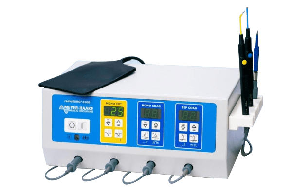
The radioSURG®2200 operates at a frequency of 2.2 MHz, which minimizes tissue heating in the incision area. As a result, there are practically no changes at the cellular level, and the tissue sample can be used for histological examination up to its edges.
The advantage of radiosurgery when used correctly:
Rapid wound healing;
Ease of operation without the need for pressure on the tool;
Sterile operating field;
No major bleeding.

radioSURG®2200 features:
3 waveforms (fully filtered wave, fully rectified wave and partially rectified wave);
Adjustable degree of coagulation;
Adjustable coagulation pulse duration from 0.5 to 0.45 seconds;
Cutting is done without tissue tension and pressure, which is especially important with the most even and accurate cut;
Minimal postoperative patient discomfort;
Minimizes the likelihood of cicatricial complications;
Tissue regeneration occurs at maximum speed;
The radio wave has a sterilizing effect and the electrode is always sterile;
When carrying out coagulation, the radio wave allows for the minimum time to transfer the maximum energy to the vessel and brew it without causing deep necrosis;
All electrodes are reusable.

The RadioSURG 2200 device is actively used in various fields of medicine:
general surgery;
ophthalmology;
vascular surgery;
gynecology;
pediatrics;
dental and maxillofacial surgery;
oncology;
neurosurgery;
cosmetology;
plastic and reconstructive surgery.
Operating modes of the radioSURG 2200 radiosurgical device
incision (power up to 100 W contributes to the achievement of an excellent cosmetic effect, the mode can be used for biopsy);
incision and coagulation (9 steps of power distribution from 10 to 90%, sparing effect on tissues);
coagulation (pulse (0.05 - 0.45 sec) and continuous modes of operation, adjustable power up to 90W, ensuring hemostasis or destruction of mucous membranes and tissues);
bipolar coagulation (makes it possible to coagulate a separate vessel, performed with special tweezers, pulsed or constant mode, power and degree of coagulation are adjustable).

Advantages of radioSURG 2200
The presence of separate outputs for the incision electrode, incision with coagulation and coagulation electrode with auto-activation allows you to work without resorting to replacing the electrode in the handle.
Re-adjustment of the electrode is not required - the device remembers the set operating parameters.
The radioSURG 2200 can be mounted on a convenient transport trolley with a smoke aspirator.
Synchronous operation of radioSURG 2200 and a smoke aspirator allows:
– regulate the speed of aspiration;
- effectively remove smoke, which greatly facilitates the work of the surgeon;
– adjust the operating time of the smoke aspirator after turning off the radioSURG 2200 in the range from 5 to 120 seconds;
– perform effective three-stage filtration for particles whose diameter is less than 0.014 microns (removal of odor, steam, volatile smoke particles, viruses);
– simple replacement of filters (resource from 3 to 6 months);
– maximum smoke suction power 990l/min.
MICROSCOPE NEUROSURGICAL LEICA M220 F12
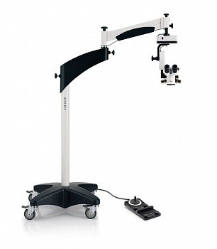
Economically attractive, the Leica M220 fully meets the needs of everyday eye surgery. With superior optics, innovative LED lighting and intuitive, user-friendly controls, the Leica M220 F12 can be a trusted “friend” to the surgeon, relieving physician fatigue and providing peace of mind for years to come.

Main advantages:
– Motorized apochromatic 5-step zoom change and focus adjustment for maximum productivity.
– Superb Leica optics produce crisp, high-contrast images with excellent depth of field for fatigue-free working.
– Instant and absolutely stable red reflection thanks to the unique direct LED light source.
– No fiber optic cable and LED elements with a rated life of 60,000 hours provide many years of reliable operation at minimal cost.
– Compact dimensions, large extension of the carrier rod provide a high degree of freedom in placing the microscope.
– Leica quality: best optics, high precision, reliability.
FOROPTOR HUVITZ HDR-9000
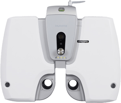
- Ophthalmic phoropter
- The latest, modern electronic phoroptor HDR-9000 from Huvitz. Advanced technology, modern design.
- When developing the HDR-9000 phoroptor, the wishes and recommendations of many optometrists from around the world were taken into account.
- Standard study program
The new program contains 21 steps, the program allows a specialist who does not have much experience to conduct a patient examination quickly and accurately. All research results are displayed on the control panel display and are available to both the doctor and the patient. - Cross Cylinder
A double cross-cylinder, as well as a cross-cylinder Jackson are used to refine the axis of the cylinder. The increased lens change speed prevents accommodation from being triggered and guarantees accurate results. - Monocular eye height adjustment
Individual for each eye adjustment of the height of the level in the range: + / — 3 mm. - Body tilt function
The latest feature, when testing near vision, allows you to tilt the body of the device from 0° to 45°, providing the feeling that the patient is reading a book. - Compatible with sign screen projectors
Phoroptor HDR-9000 can work in conjunction with the polarizing panels of screen projectors from various manufacturers. - Fast and silent lens change
Quick and quiet lens changes help minimize the effects of accommodation. Silent lens change raises the level of comfort during patient testing. The thinner body design prevents the “tunnel effect” during the examination. - Diagnostic tables
Tests for determining near visual acuity, implemented through high quality charts. The device contains additional studies for testing visual functions. - On-line assistant
On-screen graphical representation assists in the real-time testing process. With the help of hints, the research process is faster and easier. - Additional tests
The HDR-9000 phoroptor program includes a wide range of diagnostic studies, such as a color perception test, Amsler grid and other types of tables designed to check the parameters of vision at a close distance. - Tilt-and-turn display
Regardless of the patient’s position, the information on the control panel display is easy to recognize. - Built-in printer
The thermal printer built into the control panel provides fast and convenient printing of research results. - Monocular PD adjustment
Many patients need to adjust PD monocularly, the HDR-9000 Phoroptor provides independent PD adjustment for each eye. - Automatic convergence function
When the near visual acuity test is enabled, the automatic convergence function forces the test subject to look at the test chart through the center of the phoroptor lens, thereby providing a more accurate examination. - Illumination of the vertex distance check window
A more accurate examination is guaranteed by positioning the patient’s eye, the correct position of the patient’s eyes is controlled through a special illuminated window on the device body. - Lighting / Removable near table
The built-in LED table lighting for near recognizes whether the near test mode is on or off, thereby creating a comfortable table lighting. The near table is easily removed after the examination is completed. - Displaying the result in tables and graphs
The results of the patient’s studies are presented in tables and graphs, which makes it easier and faster to analyze the data obtained. - Wireless connection
Wireless communication with HRK-9000A and HLM-9000 is via Wi-Fi, previous models are connected via RS-232. - Tablet control (option)
The HDR-9000 phoroptor can be controlled not only from the control panel, it is also possible to use a tablet computer as a control console. Tablet PC OS Win7/Win8/Screen Resolution 1366*768
FOROPTOR HUVITZ HDR-7000
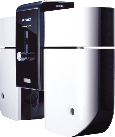
- Automatic phoropter, double cross cylinder
- Designed for the most comfortable determination of subjective refraction, selection of glasses and contact lenses. A single operating panel allows you to control the phoroptor and the sign projector, transfer objective refraction data from the HRK-7000 autorefractometer and spectacle lens parameters from the HLM-7000 dioptrimeter to the phoroptor.
- Automation of all measurement processes ensures accurate results with minimal effort!
- On-line explorer
The process of checking vision and fitting lenses becomes faster and easier thanks to the graphical representation of the tests. - Additional tests
The program of the automatic phoroptor contains a wide range of diagnostic methods, such as a test for color blindness, the Amsler Grid and other types of charts designed to check the parameters of vision at a close distance. - Double cross cylinder
The double cross cylinder is very handy for doing a «grit» test to refine force and axis. The built-in prism divides the field of view, allowing the subject to simultaneously see and compare two images. - Touch screen
The intuitive interface of the electronic phoroptor provides the operator with maximum comfort in work. The touch screen with conveniently located buttons allows you to start the necessary measurements with one touch.