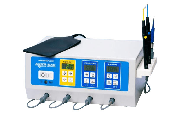
The radioSURG®2200 operates at a frequency of 2.2 MHz, which minimizes tissue heating in the incision area. As a result, there are practically no changes at the cellular level, and the tissue sample can be used for histological examination up to its edges.
The advantage of radiosurgery when used correctly:
Rapid wound healing;
Ease of operation without the need for pressure on the tool;
Sterile operating field;
No major bleeding.

radioSURG®2200 features:
3 waveforms (fully filtered wave, fully rectified wave and partially rectified wave);
Adjustable degree of coagulation;
Adjustable coagulation pulse duration from 0.5 to 0.45 seconds;
Cutting is done without tissue tension and pressure, which is especially important with the most even and accurate cut;
Minimal postoperative patient discomfort;
Minimizes the likelihood of cicatricial complications;
Tissue regeneration occurs at maximum speed;
The radio wave has a sterilizing effect and the electrode is always sterile;
When carrying out coagulation, the radio wave allows for the minimum time to transfer the maximum energy to the vessel and brew it without causing deep necrosis;
All electrodes are reusable.

The RadioSURG 2200 device is actively used in various fields of medicine:
general surgery;
ophthalmology;
vascular surgery;
gynecology;
pediatrics;
dental and maxillofacial surgery;
oncology;
neurosurgery;
cosmetology;
plastic and reconstructive surgery.
Operating modes of the radioSURG 2200 radiosurgical device
incision (power up to 100 W contributes to the achievement of an excellent cosmetic effect, the mode can be used for biopsy);
incision and coagulation (9 steps of power distribution from 10 to 90%, sparing effect on tissues);
coagulation (pulse (0.05 - 0.45 sec) and continuous modes of operation, adjustable power up to 90W, ensuring hemostasis or destruction of mucous membranes and tissues);
bipolar coagulation (makes it possible to coagulate a separate vessel, performed with special tweezers, pulsed or constant mode, power and degree of coagulation are adjustable).

Advantages of radioSURG 2200
The presence of separate outputs for the incision electrode, incision with coagulation and coagulation electrode with auto-activation allows you to work without resorting to replacing the electrode in the handle.
Re-adjustment of the electrode is not required - the device remembers the set operating parameters.
The radioSURG 2200 can be mounted on a convenient transport trolley with a smoke aspirator.
Synchronous operation of radioSURG 2200 and a smoke aspirator allows:
– regulate the speed of aspiration;
- effectively remove smoke, which greatly facilitates the work of the surgeon;
– adjust the operating time of the smoke aspirator after turning off the radioSURG 2200 in the range from 5 to 120 seconds;
– perform effective three-stage filtration for particles whose diameter is less than 0.014 microns (removal of odor, steam, volatile smoke particles, viruses);
– simple replacement of filters (resource from 3 to 6 months);
– maximum smoke suction power 990l/min.
Метка: Neurosurgery
MICROSCOPE NEUROSURGICAL LEICA M220 F12
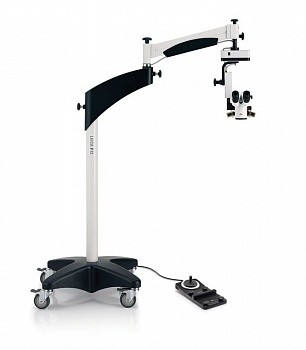
Economically attractive, the Leica M220 fully meets the needs of everyday eye surgery. With superior optics, innovative LED lighting and intuitive, user-friendly controls, the Leica M220 F12 can be a trusted “friend” to the surgeon, relieving physician fatigue and providing peace of mind for years to come.

Main advantages:
– Motorized apochromatic 5-step zoom change and focus adjustment for maximum productivity.
– Superb Leica optics produce crisp, high-contrast images with excellent depth of field for fatigue-free working.
– Instant and absolutely stable red reflection thanks to the unique direct LED light source.
– No fiber optic cable and LED elements with a rated life of 60,000 hours provide many years of reliable operation at minimal cost.
– Compact dimensions, large extension of the carrier rod provide a high degree of freedom in placing the microscope.
– Leica quality: best optics, high precision, reliability.
MICROSCOPE NEUROSURGICAL HAAG-STREIT SURGICAL HI-R

The premium microsurgical system is based on a Hi-R microscope and the latest FS 5-33 (or FS 3-43) floor stand. The system has a number of unique features and characteristics that can surprise many experienced neurosurgeons. The optical properties of the microscope meet the highest quality standards.
Apochromatic optics provide accurate color reproduction, sharp contrast and high resolution. The impressively wide stereobase (25 mm distance between the optical channels of the microscope) provides an excellent three-dimensional image in the eyepieces and enhances the surgeon's spatial perception of the operating field and instruments.

Peculiarities
Unique SensoServo technology keeps the microscope in constant balance
Depth of field control
Aperture against blinding light
New long arm tripod with innovative vibration reduction technology
Modular system design with full integration of all modules
Support for navigation stations
Computer control of all motorized functions
Optional: ICG fluorescein angiography and ALA neuro-oncology module
Optional: high-resolution image injection
Optional: World's first system with iOCT optical coherence tomography

Modular design
The new generation of tripods, which are used in the creation of neurosurgical systems, have a wide range of options for equipping with additional modules. The microscope may include a light source, a video camera and registration system, a 3D full HD video system, additional light sources, an iOCT intraoperative optical tomograph processing module, and much more. All equipment is mounted on a tripod and becomes "one" with the microscope. Such a complex system is controlled by the C.TAB touch display, which is reminiscent of today's tablets and smartphones in style and feel. The diagonal of the C.TAB display is 10.4 inches.

The world's first operating microscope with iOCT optical coherence tomography
The method is based on optical scanning, the penetration depth of which is from 0.5 to 3 mm, depending on the density of the tissue. In real time, the surgeon sees slices in the region of interest (as if in vivo histological slices), which are visualized on a small display above the eyepieces or projected directly into the surgeon's eyepieces (Image Injection).

Unsurpassed positioning system
The unique SensoServo technology provides unsurpassed positioning accuracy of the microscope in space with no effort on the part of the surgeon. The degree of applied pressure on the handles is recorded by built-in sensors that control the electric drives of the microscope and provide accurate and fast response to commands. The inertia and sensitivity of the motors is adjusted individually. At the same time, the microscope always maintains balance, even after changing accessories or changing the position of the microscope.

Eyepiece projection (Image Injection)
HAAG-STREIT SURGICAL has developed a new eyepiece projection system with very high image quality and realistic color reproduction. The resolution of the internal display is 1280x1024 at 24-bit color depth. External signal sources are connected via common DVI or VGA interfaces. The projection works both in the overlay mode (a translucent image against the background of the operating field of the microscope) and in the full image replacement mode. Data from the navigation station, images from CT, MRI, endoscopic racks, intraoperative optical coherence tomography iOCT, for the first time combined with a microscope, can be transmitted to the eyepieces.

Intraoperative imaging
The microscope can be equipped with optional modules for neuro-oncology ALA and fluorescein angiography ICG. ALA is used in neurosurgery to visualize late stages of glioblastoma (stages III and IV), as well as areas adjacent to the tumor involved in the pathological process.
This is possible because the last step of heme synthesis is suppressed in such tumor cells, which leads to the accumulation of protoporphyrin IX (PPIX) in tumor cells. When illuminated with blue light, protoporphyrin IX emits intense red light (fluorescence) and thus highlights the underlying tumor and adjacent areas, which stain orange-pink.

Floor stands
The microsurgical system can be assembled on various tripods, which differ in shoulder extension, bearing capacity and price. For example, if you need a full-featured system for spinal and neurosurgery without special fluorescence imaging tools, the FS 3-43 tripod microscope is ideal. If it is necessary to equip the microscope with all possible modules and functions, the manufacturer recommends the latest tripod FS 5-33 with an innovative vibration suppression system and a very long shoulder extension.
MICROSCOPE NEUROSURGERY CARL ZEISS ZEISS OPMI VARIO 700

ZEISS OPMI Vario 700 is a universal modular operating microscope for spinal surgery, plastic and reconstructive surgery, as well as neurosurgical and otolaryngological operations.
OPMI VARIO 700 provides the highest quality display of any parts of the working surface without chromatic aberrations and geometric distortion of the image.
apochromatic optics with multilayer antireflex coating
optimal visualization in the range of 200–500 mm.
180/300W fiber optic xenon lighting
automatic diaphragm.
OPMI VARIO 700 is equipped with a fully integrated 3CCD video camera and image recording system. This allows surgeons to capture very clear images without additional equipment. Images can be broadcast on the web, stored in a patient's medical record, or used for presentations and subsequent analysis.
The OPMI VARIO 700 configuration includes a 170/260mm expandable swivel tube with 12.5x wide-angle magnetically coupled plug-in eyepieces. It allows you to quickly switch distances, gives an additional increase of 50% and is easily fixed in any convenient position.
The microscope has a simple and intuitive interface that speeds up preoperative setup through simplified access to all microscope functions. Programmable knobs, a pendant display and a multi-functional foot control provide comfort during surgery.
OPMI VARIO 700 is equipped with a full auto-balancing system at the touch of a button on the touch control panel, an auto-shroud system and monitoring the life of the lighting system.
MICROSCOPE NEUROSURGICAL CARL ZEISS ZEISS OPMI VARIO S88
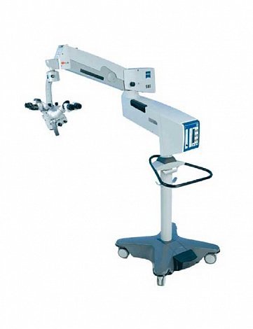
ZEISS OPMI Vario S88 is a universal compact operating microscope for ENT - microsurgery, neuro and spinal, vascular, plastic, reconstructive surgery and hand microsurgery. It provides uniform illumination and perfect detail of fine structures even in narrow and deep canals.
ZEISS OPMI Vario S88 allows the surgeon to take a comfortable position even when performing operations on the base of the skull. The microscope can be controlled using the multifunctional foot control or the touch control panel on the tripod. It is equipped with electromagnetic clutches that fix the suspension system in the desired position and allow you to effortlessly move the microscope with one hand.
The microscope allows you to take photo and video equipment, record, archive and edit video images, display images on a monitor. Apochromatic optics with multilayer anti-reflective coating, xenon illuminator and 1:6 zoom provide improved image quality, high resolution and color accuracy.
ZEISS OPMI Vario S88 gives both the doctor and the assistant the opportunity to work with magnification. An assistant microscope, located at an angle of 90 degrees from the main surgeon's tube, allows you to see the entire surgical field and perform many hours of surgery.
MICROSCOPE NEUROSURGICAL CARL ZEISS ZEISS OPMI PICO ENT

ZEISS OPMI Pico ENT is a high quality system with a compact and robust design. Designed for everyday use in the practice of an otorhinolaryngologist. The latest optics, combined with advanced imaging technologies such as the built-in HD video chain, give you and your patient a clear view of what is happening. Main characteristics LED lights This light source is virtually maintenance-free and allows you to see details as clearly as in daylight. Optics ZEISS Varioskop Once a Varioskop® lens is attached, the ZEISS OPMI pico system allows you to seamlessly adjust the focal length to suit your targets - ear, nose or throat - simply by turning a knob. Combined with one of the S100 suspension systems: floor stand, ceiling or floor bracket, the ZEISS OPMI pico can be easily adapted to virtually any work environment. Integrated Video Solutions Comprehensive video solutions for operating microscopes are the most convenient way to create images for informational, documentary, educational and presentation purposes. With built-in camera systems with factory-tuned optics, these video solutions can be up and running right out of the box. Built-in full HD video camera ZEISS OPMI pico is equipped with a built-in HD video camera with live streaming and a video recording license. The chamber is pre-built with optimized and proven settings for the work of an otorhinolaryngologist. Video frames and images can be displayed on a video monitor using standard video interfaces, or broadcast over the network online for viewing on various devices or for complex video recording. The smart recording function works in retroactive mode, recording data up to 30 seconds before the start of shooting. Halogen (left) and LED (right) illumination The LED backlight provides even, near-natural illumination that allows you to clearly see fine structures and details. The long life of the LED light sources reduces system operating costs. Varioscope 100 The Varioskop® 100 from ZEISS makes it easy for you to focus on your area of interest. Focusing on the object is carried out by simply turning the knob located on the microscope objective. Smoothly adjust the focal length you need in the range of 200-300 mm without changing lenses. Protective lenses VisionGuard ZEISS Surgical Drapes are manufactured with ZEISS's unique VisionGuard® Optical Lens, which works together with the surgical microscope objective as a single optical instrument, providing optical clarity. During surgery, the lens can be replaced with a new sterile lens without compromising sterility. OPMI pico microscope with endoscopic camera port Attaching endoscopic cameras Quickly attach an existing endoscopic camera to the ZEISS OPMI pico microscope. OPMI pico microscope on wall bracket Adjustable to suit your workspace The ZEISS OPMI pico is available with a variety of systems: the S100 can be floor-mounted, ceiling-mounted, or wall-mounted. It can be built into the workplace of an ENT doctor.
MICROSCOPE NEUROSURGICAL CARL ZEISS ZEISS OPMI PENTERO 900
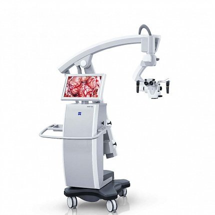
ZEISS OPMI PENTERO® 900 provides superior visualization of the surgical process thanks to apochromatic optics and innovative intraoperative fluorescence modules. It also allows you to record full HD video images.
The system is controlled using the touch screen, handles, mouthpiece or wireless foot control. Incredible functionality thanks to the extendable tube, f170/f260.
HD video solutions ZEISS OPMI PENTERO allows you to quickly and easily create images for informational, documentary, educational and presentation purposes. Comprehensive control allows the organization of the video process together with the microscope, thereby contributing to a more coordinated work of surgeons.

Main characteristics


High-contrast, sharp images with integrated high-definition 3-smart HD camera (1080p)
Video images with optimized clarity and color for high-end surgical imaging through a specially designed camera system for the ZEISS OPMI PENTERO 900
The video camera's light sensitivity provides sharp, high-contrast images for intraoperative use using fluorescent illumination (BLUE™ 400 and YELLOW™ 560)
Operation of the integrated high-definition (1080i) HD video recorder can be performed via touch screen, wireless foot control or operating microscope handle
Videos and images are stored on the integrated hard drive and can also be transferred directly to USB sticks or to a shared LAN directory
Memory capacity can be optimized by using the local editing function. Large HD video footage can be edited quickly and easily on the operating microscope's touch screen
Exchange of standardized data with image archiving and communication systems (PACS) through the integrated module for the transmission and visualization of digital medical images and documents of examined patients (DICOM)
The integrated 22" monitor provides excellent image quality of the surgical field. The operating room team can comfortably observe the progress of the operation due to the wide viewing angle and flexible arm of the monitor
Images from the integrated video device can be displayed on secondary monitors or other video systems in the operating room using standard video interfaces.

Features and functions

Intraoperative fluorescence module ZEISS INFRARED 800
Intraoperative visual assessment of blood flow and vascular patency during surgery, shunting and aneurysm removal.

Intraoperative fluorescent module FLOW 800
A unique program that allows you to visually analyze the vascular dynamics of blood flow. It assembles sequences of INFRARED 800 video frames into visual maps, diagrams, or side-by-side images, allowing in-depth interpretation of video frames captured under fluorescent light.

Intraoperative fluorescent module YELLOW 560
YELLOW™ 560 provides imaging with fluorescent dyes with a wavelength of 540-690 nm. This is the first fluorescence module from ZEISS that allows the visualization of fluorescein-stained structures against a background of unstained tissue. It is also available in HD quality.

Intraoperative Fluorescence Module BLUE 400
BLUE™ 400 provides intraoperative identification of high grade gliomas. This is a fully integrated fluorescence module built into the microscope as a result of a successful comprehensive clinical study.

DICOM and MultiVision modules
The ZEISS OPMI PENTERO 900 offers a comprehensive connectivity configuration with workspace technology and data management functionality. Integrated modules such as MultiVision™ and DICOM enable seamless communication with other imaging devices and therefore connection to a specific hospital's communications infrastructure.

Sliding tube f170 f260
The flexible folding tube f170/f260 features improved positioning, magnification and user comfort. With a quick turn of the adjustment knob, the integrated PROMAG™ functionality provides an additional 50% magnification.

ZEISS 3-stage zoom switch
Provides additional magnification when submillimeter imaging is required using the ZEISS 3-stage magnification switch.

Shared Surveillance Stereo Tube
The co-observation stereotube allows the second surgeon to see the surgical field at the same magnification as the first surgeon. This is especially useful for assistants helping with sterilization or training.

Sterilizable sheaths with protective lens Visionguard
ZEISS Surgical Drapes are manufactured with ZEISS's unique VisionGuard® Optical Lens, which works together with the surgical microscope objective as a single optical instrument, providing optical clarity. During surgery, the lens can be replaced with a new sterile lens without compromising sterility.

Wireless foot control
Designed to control the many functions of the operating microscope, the wireless foot controller can be freely positioned for quick setup and user convenience during surgery. The intelligent power management feature ensures long-term serviceability.
MICROSCOPE NEUROSURGERY CARL ZEISS ZEISS OPMI PENTERO 800
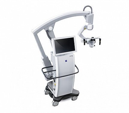
ZEISS OPMI PENTERO 800 is a state-of-the-art neurosurgery operating system that enables critical operations on fine structures through deep channels. Thanks to apochromatic optics and unsurpassed ergonomics, the operating microscope provides excellent image clarity and optimal operating efficiency.
The ZEISS OPMI PENTERO 800 features advanced apochromatic optics, dual xenon illumination and a fully integrated video recording system. This allows the doctor to see the smallest details of the operation on the monitor or through the eyepieces.
The system has a user-friendly user interface that provides direct access to all operating microscope functions via the touch screen. This helps to quickly obtain information about the patient and adjust device settings.
ZEISS OPMI PENTERO 800 is comfortable to use and ergonomic. The microscope's floating arm and dynamic folding tube provide maximum comfort and greatly expand magnification and positioning options.
MICROSCOPE NEUROSURGICAL CARL ZEISS ZEISS OPMI MOVENA
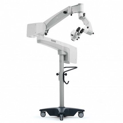
ZEISS OPMI Movena is a compact, manoeuvrable otolaryngology operating microscope with varioscope and spotlight.
The surgeon can move the OPMI Movena with just one movement of the head. At the same time, the balancing system provides amazing ease of movement, and the microscope will maintain positioning accuracy.
The built-in manual zoom switch, combined with the manual focus system, allows you to select any desired working distance from 200mm to 415mm without changing lenses. An external fine focusing system ensures the alignment of the laser and optical beam, the highest focusing accuracy when working with an attached laser micromanipulator.
Aiming lighting system allows you to reduce the diameter of the light spot to 11 mm. The surgeon can set the desired intensity of illumination using a conveniently located brightness control knob.
OPMI® Movena™ has a compact design that fits perfectly into any space. The small dimensions of the microscope give great advantages to surgeons performing interventions on the larynx and allow active use of additional features during the operation.
The OPMI® Movena™ microscope can be equipped with the S7 floor stand or the S7 ceiling mount:
The S7 floor stand makes it easy to move the microscope during surgery, as well as transport it between operating rooms.
The S7 ceiling mount with fixed or adjustable column height keeps the microscope within easy reach.
MICROSCOPE NEUROSURGICAL CARL ZEISS OPMI SENSERA
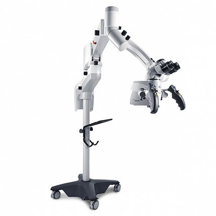
ZEISS OPMI Sensera is a compact operating microscope designed for delicate and precise manipulations on the ear and skull base. Its distinctive feature is the presence of an internal focusing mechanism, a varioscope.
OPMI Sensera allows you to clearly see the smallest structures in natural colors with Superlux180 xenon illumination and a focus range of 200-415mm.
The complete set of the OPMI Sensera microscope makes it possible to conveniently and efficiently perform any surgical procedures:
Compact design to fit in any size operating room
Binocular swivel tube 180°
Spring balancing system for quick and easy microscope balancing even when using the maximum number of accessories.

Integrated video camera, the ability to combine with a video recording system and a medical monitor.
The OPMI Sensera microscope can be easily and quickly adapted to the needs of the operating room. In addition, personal settings for up to 9 users can be saved.